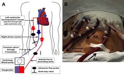
lv vent | left ventricular venting lv vent Venoarterial extracorporeal membrane oxygenation (VA ECMO) is an established method of short-term mechanical support for patients in cardiogenic shock, but can create left ventricular . Ejection fraction (EF) is a measurement, expressed as a percentage, of how much blood the left ventricle pumps out with each contraction. An ejection fraction of 60 percent means that 60 percent of the total amount of blood in the left ventricle is pushed out with each heartbeat.
0 · left ventricular venting
1 · left ventricular unloading
2 · left ventricular hypertrophy treatment
3 · Lv vent surgery
4 · Lv vent procedure pdf
5 · Lv vent procedure
6 · Lv vent cardiac surgery
7 · Lv vent cannula
A condensation of Shimano's latest technologies is the Antares DC, giving you the casting distance you need by incorporating the MGL Spool III onto a DC system (the first of its kind!). Coupled with the fine-tuned 4x8 DC brake system, a multiplier effect can be seen when it comes to casting far distances.
Venoarterial extracorporeal membrane oxygenation (VA ECMO) is an established method of short-term mechanical support for patients in cardiogenic shock, but can create left ventricular .Surgical LV vent. Surgical Technique – insertion of apical left ventricular vent. by Prof David McGiffin. Preparation. The patient is placed with a 30-degree bump on the left side. The .
LV venting is an important adjunct of myocardial protection during systemic cooling before successful delivery of cardioplegia. Conventionally, LV vent is placed via the right superior .Indications to vent the LV are variable and can be based on clinical, hemodynamic, or echocardiographic findings of impaired LV unloading, LV stasis, or pulmonary edema. Indeed, . I'm trying to find the correct coding for insertion of a left ventricular vent only (not an assist device) placed on a date after Ecmo cannulation (36822) on a pediatric patient on . Good Morning, I am wondering if I coded this note correctly: INDICATIONS: A 66 year old man with history of severe cardiomyopathy and congestive heart failure. He .
The descriptor for CPT 33244 doesn't specify where the leads are removed from (LV, RV, RA), only that it is by transvenous extraction so it is the correct code for removal of . The LV vent was removed and that site closed with the previously placed pursestring. Echocardiography was then used to interrogate the valve, which showed a low . Tourniquets were placed around both caval cannulas. An LV vent was placed through the right superior pulmonary vein. An antegrade cardioplegia catheter was placed in .
MANAGEMENT OF AIR: Patient was in Trendelenburg. LV vent was turned off. The left heart was filled. The lungs were fully inflated. Heart was vigorously massaged. Root . approximately every 20-30 minutes. An LV vent was placed through the right superior pulmonary vein. I first proceeded to open the proximal aorta through a transverse . An LV vent was placed through the right superior pulmonary vein. An antegrade cardioplegic catheter was placed in the proximal ascending aorta. The aorta was then .

left ventricular venting
Best answers. 0. Mar 21, 2022. #1. PROCEDURES: Right femoral and arterial cannulation using Heartport access, right mini. thoracotomy, this would be considered a redo mediastinum to . pledgeted sutures. An LV vent was placed through the right superior pulmonary vein. The valve appeared to have much better coaptation. I therefore proceeded to close the .
I'm trying to find the correct coding for insertion of a left ventricular vent only (not an assist device) placed on a date after Ecmo cannulation (36822) on a pediatric patient on .
Good Morning, I am wondering if I coded this note correctly: INDICATIONS: A 66 year old man with history of severe cardiomyopathy and congestive heart failure. He .
The descriptor for CPT 33244 doesn't specify where the leads are removed from (LV, RV, RA), only that it is by transvenous extraction so it is the correct code for removal of .
The LV vent was removed and that site closed with the previously placed pursestring. Echocardiography was then used to interrogate the valve, which showed a low . Tourniquets were placed around both caval cannulas. An LV vent was placed through the right superior pulmonary vein. An antegrade cardioplegia catheter was placed in . MANAGEMENT OF AIR: Patient was in Trendelenburg. LV vent was turned off. The left heart was filled. The lungs were fully inflated. Heart was vigorously massaged. Root .
approximately every 20-30 minutes. An LV vent was placed through the right superior pulmonary vein. I first proceeded to open the proximal aorta through a transverse . An LV vent was placed through the right superior pulmonary vein. An antegrade cardioplegic catheter was placed in the proximal ascending aorta. The aorta was then .Best answers. 0. Mar 21, 2022. #1. PROCEDURES: Right femoral and arterial cannulation using Heartport access, right mini. thoracotomy, this would be considered a redo mediastinum to .
left ventricular unloading
richard mille watch odell
richard mille watch toronto
richard mille watch michelle yeoh
left ventricular hypertrophy treatment
Material Name: Flexible FAST Adhesive Part B Synonyms: Polyol Blend. Chemical Family: Polyol Blend Product Use: Commercial Roofing Adhesive Restrictions on Use: Professional use only Manufacturer Information Carlisle SynTec 1285 Ritner Highway Carlisle, PA 17013 USA Phone: +1-800-479-6832 Emergency Phone #: +1-800-424-9300 (CHEMTREC)
lv vent|left ventricular venting


